
Diagnosing Atopic Dermatitis
The signs and symptoms
AD is diagnosed clinically based on medical history, morphology and distribution of skin lesions, and associated clinical features. When examining patients, the signs and symptoms of AD to look out for are1:
Essential
(must be present)
- Pruritus (itch).1
- Acute, subacute, or chronic eczema.1
Important
(observed in most cases)
- Early age of onset.1
- Atopy with personal and/or family history and immunoglobulin E reactivity1
- Xerosis.1
Associated
(non-specific, helps suggest a diagnosis of AD)
- Atypical vascular responses (eg, facial pallor, white dermographism, and delayed blanch response).1
- Keratosis pilaris/pityriasis alba/hyperlinear palms/ichthyosis.1
- Ocular/periorbital changes.1
- Other regional findings (eg, perioral changes/periauricular lesions).1
- Perifollicular accentuation/lichenification/prurigo lesions.1
- Associated conditions and clinical associations such as allergies, asthma, allergic rhinitis/rhinoconjunctivitis, sleep disturbance, depression, cancer, and obesity.1

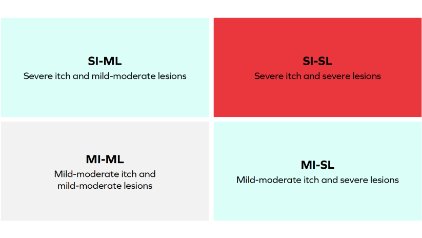
Patients With AD May Present With Different Phenotypes
AD is a heterogeneous disease associated with multiple phenotypes and variable intensity of itch and lesions.5
Itch-dominant (SI-ML) AD is a common, burdensome, and distinct subtype of AD, occurring in up to 29% of patients.
The proportion of patients with itch-dominant AD was higher in women and patients of African descent.5

Differentiating AD from PN
AD presents with an often diffuse distribution of eczematous lesions while PN presents with an often symmetrical distribution of firm, nodular lesions.2-4

The damaging disease burden
Persistent pruritus is much more than just an irritating sensation—it is the most burdensome symptom for patients with AD and can significantly disrupt their daily activities, sleep, and psychological well-being.2,6
It is recommended that you ask your patients with AD about3,7,8:
- Pruritus (itch)
- Sleep
- Impact on daily activity (including effects on work, school, and well-being)
- Disease persistence
The signs and symptoms of AD in skin of colour may differ in visual appearance, predominantly due to differences in pigmentation and lesion distribution. It is important to recognise these differences in your patients with skin of colour9:
AD in patients of colour
Pigmentation differences
- Erythema that may appear violaceous9
- Lichenification, hyperlinearity of the palms, Dennie-Morgan lines, and diffuse xerosis9
- Postinflammatory dyspigmentation (including hyperpigmentation and hypopigmentation)9
Location of lesions
- Extensor involvement, which may be more common than flexural dermatitis9
- Perifollicular accentuation and scattered distinct papules on the extensor surfaces and trunk9
Things to note
- Compared with White patients, those of Asian descent are more likely to have well-demarcated lesions and increased scaling and lichenification9
Learn More


- Eichenfield LF, Tom WL, Chamlin SL, et al. Guidelines of care for the management of atopic dermatitis: section 1. Diagnosis and assessment of atopic dermatitis. J Am Acad Dermatol. 2014;70(2):338-351. doi:10.1016/j.jaad.2013.10.0109
- McCleary KK. More than skin deep: understanding the lived experience of eczema. March 18, 2020. Accessed February 16, 2022. http://www.morethanskindeep-eczema.org/report.html
- Langan SM, Irvine AD, Weidinger S. Atopic dermatitis. Lancet. 2020;396(10247):345-360. doi:10.1016/S0140-6736(20)31286-1
- Ständer S. Atopic dermatitis. N Engl J Med. 2021;384(12):1136-1143. doi:10.1056/NEJMra2023911
- Chovatiya R, Lei D, Ahmed A, et al. Clinical phenotyping of atopic dermatitis using combined itch and lesional severity: a prospective observational study. Ann Allergy Asthma Immunol. 2021;127(1):83-90.e2. doi:10.1016/j. anai.2021.03.019
- Yaghmaie P, Koudelka CW, Simpson EL. Mental health comorbidity in patients with atopic dermatitis J Allergy Clin Immunol. 2013;131(2):428-433 doi:10.1016/j.jaci.2012.10.041.
- Janmohamed SR, Gwillim EC, Yousaf M, Patel KR, Silverberg JI. The impact of prurigo nodularis on quality of life: a systematic review and meta-analysis.Arch Dermatol Res. 2021;313(8):669-677 doi:10.1007/s00403-020-02148-0
- Todberg T, Zachariae C, Skov L. Treatment and burden of disease in a cohort of patients with prurigo nodularis: a survey-based study. Acta Derm Venereol. 2020;100(8):adv00119. doi:10.2340/00015555-3471
- Kaufman BP, Guttman-Yassky E, Alexis AF. Atopic dermatitis in diverse racial and ethnic groups-variations in epidemiology, genetics, clinical presentation and treatment. Exp Dermatol. 2018;27(4):340-357. doi:10.1111/exd.13514f
References