Drivers of Atopic Dermatitis

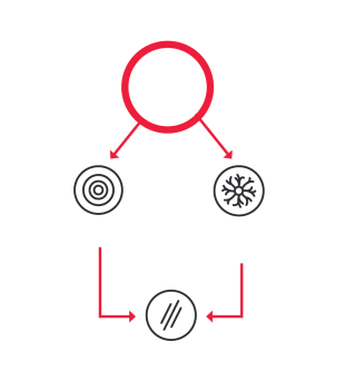
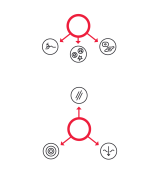
Itch, inflammation, and skin barrier disruption contribute to AD.
The neuroimmune communication between sensory neurons, keratinocytes, and inflammatory mediators (IL-4, IL-13, IL-31) contribute to AD pathophysiology.1-5

Itch is an important part of AD pathophysiology
Two pathways cause itch in patients with AD: inflammation and nerve sensitization.3,4,6-8
Inflammation
Keratinocytes, mast cells, lymphocytes, and small sensory nerve fibers in the skin release pruritogens, including IL-4, IL-13, and IL-31, inducing itch in the epidermal layers of the skin.4
Nerve sensitization
Neuroimmune cytokines, including IL-31, cause nerve elongation and branching in sensory neurons contributing to neuronal sensitization, increased sensitivity to minimal stimuli, and continuous itching.3,6-8
IL-31 has been shown to induce itching in patients with AD.
Based on human and mice studies, it has been shown that
- IL-31 induces pruritic signaling and nerve sensitization in patients with AD, leading to itch6,9,10
- IL-31 stimulates sensory neurons expressing IL-31RA to transmit pruritis signaling to the central nervous system9,10
- IL-31 stimulates neuronal growth and branching of sensory neurons, resulting in increased density of the skin neuronal network, explaining the hypersensitivity of patients with AD to itch-inducing stimuli6,9,10
- IL-31 activates keratinocytes to produce pruritis mediators, amplifying itch6,9,10
Inflammation contributes to AD pathophysiology.
Inflammatory mediators are involved in induction of inflammation in patients with AD.3-5
- IL-4 and IL-13 are involved in T cell differentiation and promote tissue inflammation, respectively3-5
- IL-31 induces inflammation via immune and nonimmune mechanisms5
IL-31 has been shown to induce inflammation in patients with AD.5,11.12
Based on human and mice studies, it has been shown that
- IL-31 induces inflammation by acting on immune and nonimmune cells7,11-13
- IL-31 activates immune cells and keratinocytes to release proinflammatory mediators, leading to skin inflammation7
- IL-31 signaling amplifies inflammation by bridging communication between epithelial cells, nerves, and immune cells7,11-13
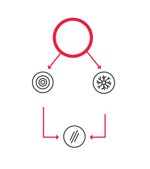
Skin barrier disruption contributes to AD.
Functional disruption of the skin barrier is attributed to filaggrin (FLG) loss-of- function mutations, inflammation, and physical damage induced by scratching.1
Cytokines IL-4, IL-13, and IL-31 are shown to disrupt skin barrier.
- IL-4 and IL-13 are associated with the decreased expression of structural proteins responsible for skin integrity, including FLG14,15
- IL-31 can directly impact FLG and further increase skin barrier disruption through inflammation14,16
IL-31 contributes to skin barrier disruption in patients with AD.9,10,17
- IL-31 overexpression impairs keratinocyte differentiation and decreases the expression of key proteins, including FLG, leading to epithelial and skin barrier disruption17
- IL-31-induced inflammation impacts skin function and aggravates barrier disruption9,10
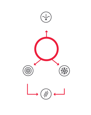
IL-31 acts as a bridge in AD, connecting nervous and immune systems and structural cells to drive AD pathophysiology.7,11-13
- Langan SM, Irvine AD, Weidinger S. Atopic dermatitis [published correction appears in Lancet. 2020;396(10253):758]. Lancet. 2020;396(10247):345-360. doi:10.1016/S0140-6736(20)31286-1
- Oetjen LK, Mack MR, Feng J, et al. Sensory neurons co-opt classical immune signaling pathways to mediate chronic itch. Cell. 2017;171(1):217-228.e13. doi:10.1016/j.cell.2017.08.006
- Mack MR, Kim BS. The itch-scratch cycle: a neuroimmune perspective. Trends Immunol. 2018;39(12):980-991. doi:10.1016/j.it.2018.10.001
- Ständer S. Atopic dermatitis. N Engl J Med. 2021;384(12):1136-1143. doi:10.1056/NEJMra2023911
- Boothe WD, Tarbox JA, Tarbox MB. Atopic dermatitis: pathophysiology. Adv Exp Med Biol. 2017;1027:21-37. doi:10.1007/978-3-319-64804-0_3
- Feld M, Garcia R, Buddenkotte J, et al. The pruritus- and TH2-associated cytokine IL-31 promotes growth of sensory nerves. J Allergy Clin Immunol.2016;138(2):500-508.e24. doi:10.1016/j.jaci.2016.02.020
- Bagci IS, Ruzicka T. IL-31: A new key player in dermatology and beyond. J Allergy Clin Immunol. 2018;141(3):858-866. doi:10.1016/j.jaci.2017.10.045
- Tominaga M, Takamori K. Peripheral itch sensitization in atopic dermatitis. Allergol Int. 2022;71(3):265-277. doi:10.1016/j.alit.2022.04.003
- Cevikbas F, Wang X, Akiyama T, et al. A sensory neuron-expressed IL-31 receptor mediates T helper cell-dependent itch: involvement of TRPV1 and TRPA1. J Allergy Clin Immunol. 2014;133(2):448-460. doi:10.1016/j.jaci.2013.10.048
- Kabashima K, Irie H. Interleukin-31 as a clinical target for pruritus treatment. Front Med (Lausanne). 2021;8:69. doi:10.3389/fmed.2021.638325
- Gibbs BF, Patsinakidis N, Raap U. Role of the pruritic cytokine IL-31 in autoimmune skin diseases. Front Immunol. 2019;10:1383. doi:10.3389/fimmu.2019.01383
- Wang F, Kim BS. Itch: a paradigm of neuroimmune crosstalk. Immunity. 2020;52(5):753-766. doi:10.1016/j.immuni.2020.04.008
- Zhang Q, Putheti P, Zhou Q, Liu Q, Gao W. Structures and biological functions of IL-31 and IL-31 receptors. Cytokine Growth Factor Rev.2008;19(5-6):347-356. doi:10.1016/j.cytogfr.2008.08.003
- Kim BE, Leung DY, Boguniewicz M, Howell MD. Loricrin and involucrin expression is down-regulated by Th2 cytokines through STAT-6. Clin Immunol. 2008;126(3):332-337. doi:10.1016/j.clim.2007.11.006
- Sehra S, Yao Y, Howell MD, et al. IL-4 regulates skin homeostasis and the predisposition toward allergic skin inflammation. J Immunol. 2010;184(6):3186-3190. doi:10.4049/jimmunol.0901860
- Cornelissen C, Marquardt Y, Czaja K, et al. IL-31 regulates differentiation and filaggrin expression in human organotypic skin models. J Allergy Clin Immunol. 2012;129(2):426–433.e4338. doi:10.1016/j.jaci.2011.10.042
- Furue M, Furue M. Interleukin-31 and pruritic skin. J Clin Med. 2021;10(9):1906. doi:10.3390/jcm10091906